Visualizing Breakthroughs, Deeper Insights

Learn How Our Animal Imaging Center Can Help
The Tufts Preclinical Imaging Center (PIC) provides equipment and expertise for in vivo and ex vivo molecular and anatomical imaging for basic research and translational studies in small animals. We offer a diverse array of imaging modalities, including optical(IVIS) imaging, ultrasound (US), micro-computed tomography (microCT), and EchoMRI. We also have a Envisu C2300 OCT, a slit lamp and Phoenix Micron IV microscope for ophthalmic studies. These modalities capture physiological and anatomical data with a wide range of spatial and temporal resolutions and imaging depths, ensuring we can support most preclinical studies. Our instruments are available both for self or technician assisted service.
The PIC not only supports research at Tufts University and affiliated hospitals, it also offers a wide range of contract research services for Biotech and Pharma. Our micro-CT is also available to materials science researchers and ex vivo work on larger animals.
Our services include: Equipment Training, Analysis Software Training, Assisted & Unassisted Imaging, Study Design/IACUC protocol consulting, and Data Post-Processing/Analysis
For confocal microscopy, please refer to the Tufts Imaging and Cell Analysis Core on the Boston Campus, and the Tufts Advanced Microscopic Imaging Center on the Medford campus.
Check Out Our Equipment Gallery
Optical Imaging (IVIS)
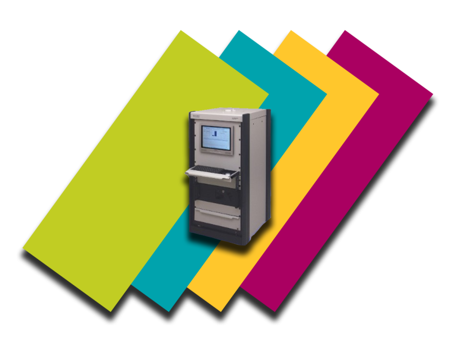
PerkinElmer IVIS Spectrum: Advanced in vivo imaging system designed for non-invasive tracking of disease progression, cell trafficking, and gene expression. Capable of bioluminescence and fluorescence imaging with high sensitivity, supporting preclinical research at Tufts CMS. Available for assisted and unassisted imaging.
Micro-Computed Tomography (microCT or uCT)
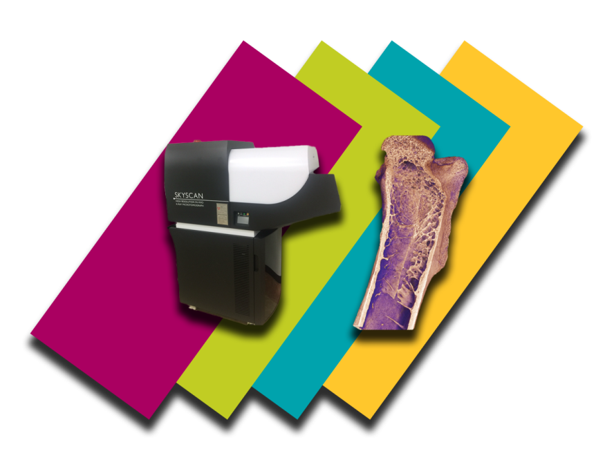
Bruker Skyscan 1176: Advanced microCT system for non-invasive volumetric imaging using X-rays. Applications include bone densitometry, morphometry, dental analysis, and lung imaging. A powerful tool for detailed cross-sectional and 3D renderings in preclinical research at Tufts CMS. Available for assisted and unassisted imaging.
Ultrasound (US)
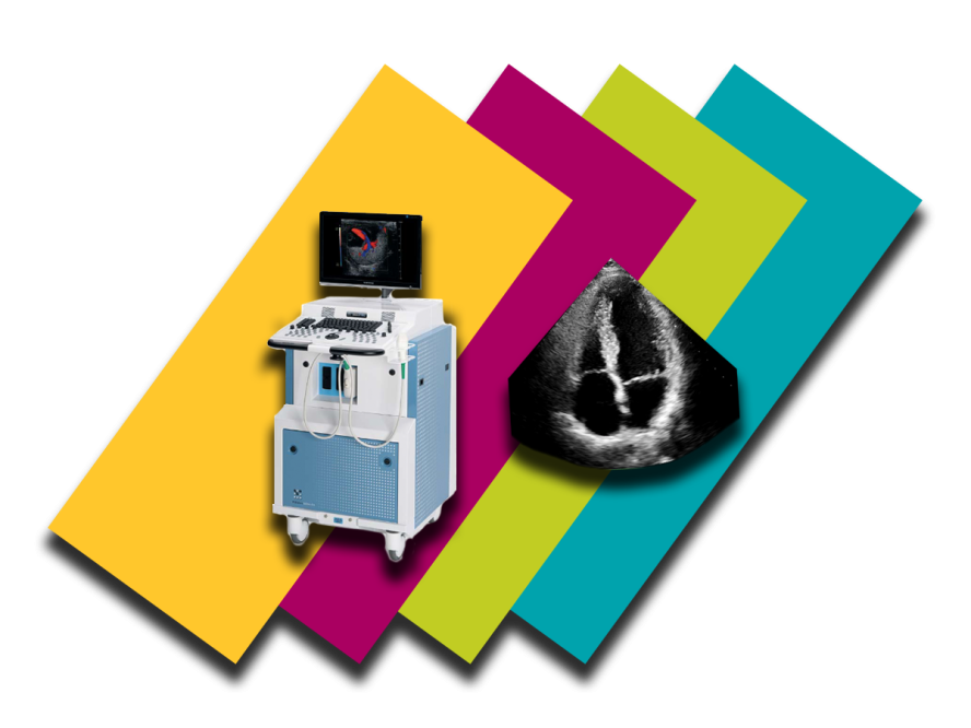
VisualSonics VEVO 2100: High-frequency ultrasound imaging system for dynamic visualization of organs, tissues, and blood flow. Ideal for applications such as Echocardiogram (Echo), cardiac function analysis, tumor monitoring, guided injections, and targeted therapy treatments. Available for assisted and unassisted imaging.
Body Composition Analyzer (BAC)
EchoMRI-100 and EchoMRI-900: Non-invasive systems for whole-body composition analysis, measuring fat, lean mass, and free water without requiring anesthesia. Essential for precise and humane preclinical studies at Tufts CMS. Available for assisted and unassisted imaging.

Ocular Coherence Tomography (OCT)
Bioptigen Envisu C2300: a spectral-domain OCT (SD-OCT) that provides high-resolution images (~3–4 µm) and fast acquisition (up to ~32,000 scans/second). A mount for rodent imaging is included.
Ocular Imaging System
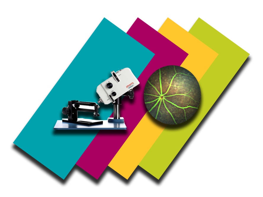
Micron IV: Advanced ophthalmic imaging system for in vivo eye and brain research. Capable of color fundus and fluorescent imaging with an image-guided laser attachment, supporting precise preclinical ophthalmic studies at Tufts CMS.
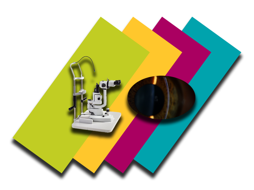
Slit Lamp
Slit Lamp: Specialized ophthalmic tool for anterior and posterior segment imaging in preclinical eye research. Provides detailed visualization of ocular structures to support advanced studies at Tufts CMS.
Preclinical Imaging Center Resources
-
We provide competitively priced, high-quality services to internal and external investigators.
Please contact us at TuftsCMS-PIC@tufts.edu to get more information about our rates and services and how we can best work with you to accomplish your research goals.
Both internal and external users can reserve imaging devices and associated procedure rooms in the Preclinical Imaging Center through iLab (an online scheduling system), after registering for an account with the site.
Review these instructions for using iLab:
-
Optical (IVIS)
- Training Video: In vitro bioluminescence imaging (BLI)
- Training Video: In vivo bioluminescence imaging (BLI)
- Training Video: In vitro fluorescence imaging (FLI)
- Training Video: In vivo fluorescence imaging (Part 1)
- Training Video: In vivo fluorescence imaging (Part 2)
- Training Video: Optical and computed tomography
MicroCT
For detailed method notes for your area of interest, please email tuftscms-pic@tufts.edu
- Training Video: 3D.Suite Overview
- Training Video: NRecon Overview
- Training Video: DataViewer Overview
- Training Video: CTan Overview
- Training Video: CTVox Overview
- Training Video: Setting a trabecular VOI
- Training Video: Setting a cortical VOI
- Training Video: BMD TMD calibration
Ultrasound
All ultrasound training videos are locked behind a password, please email tuftscms-pic@tufts.edu for access
-
Check out our FAQs' on the main FAQs Page.

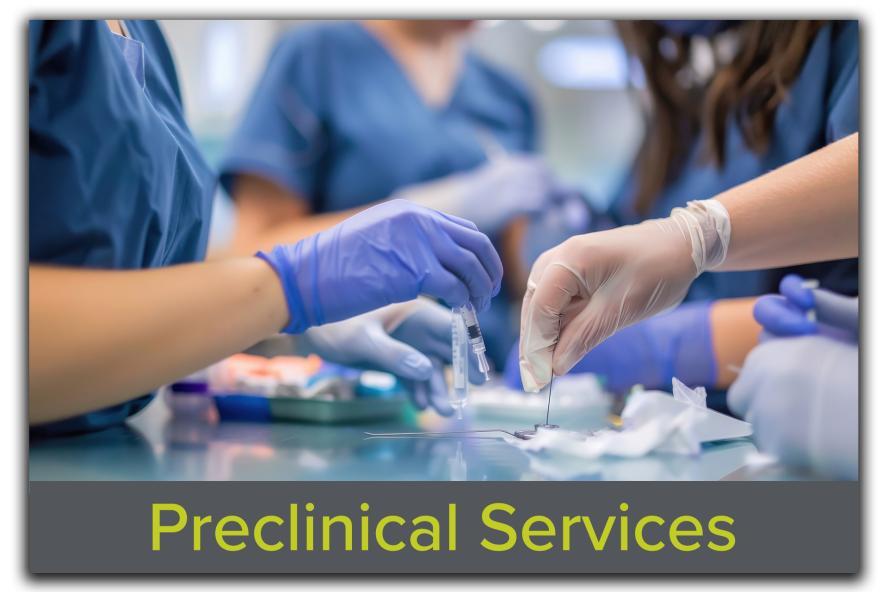
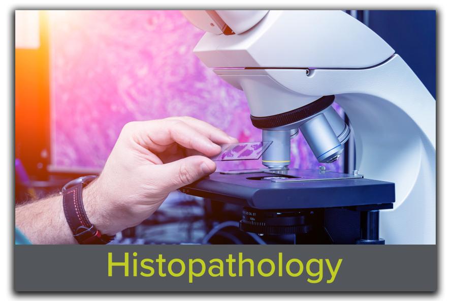
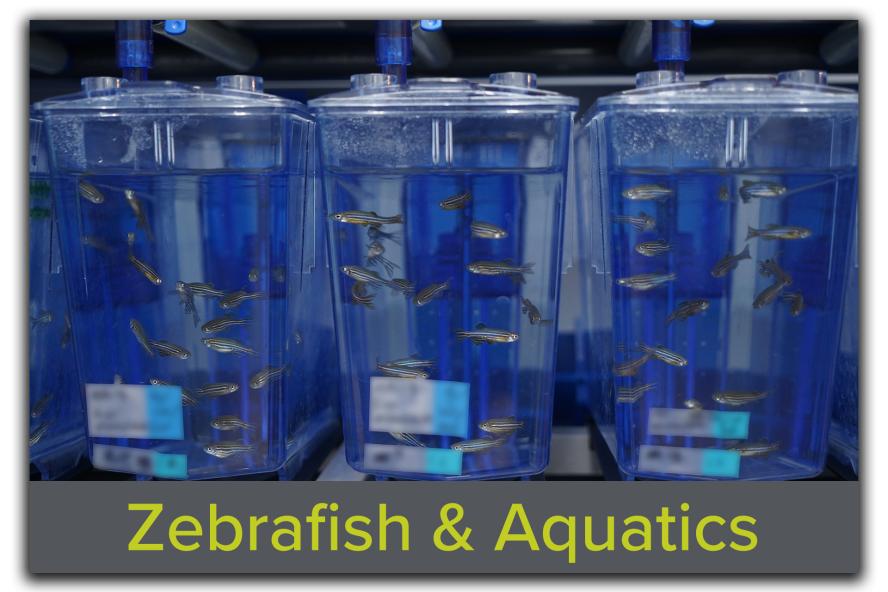
Related Services