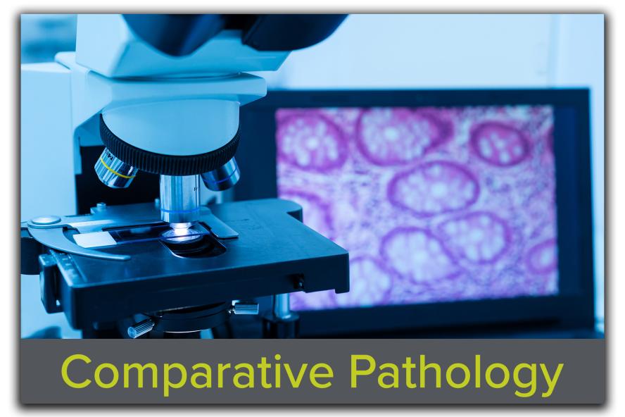
Advancing Discovery with Precision in Every Slice

Discover More About Our Histological Services
The Animal Histology Core at Tufts CMS provides full-service research animal histology support, offering tissue paraffin processing, embedding, sectioning, cryosectioning and staining techniques for research needs. Staffed by certified histology technicians, the core specializes in high-quality slide preparation and is available to consult during the planning of your study. Our histology services are designed to facilitate accurate and reproducible results for investigators across various research disciplines.
-
- Trimming of submitted fixed tissues to ensure best orientation for optimal assessment
- Decalcification of bone specimens
- Correct orientation of samples in paraffin wax
- Basic microtomy to produce standard 5µm paraffin sections
- Advanced sectioning (step sectioning, levels, thick sections, etc.)
- Unstained sections in preparation for immunohistochemistry or other procedures
-
- Preparation, freezing, and cutting of standard frozen sections
- Fixing, staining, and cover-slipping
- Both full service and self-service (after training) available.
- Self service: You may use the AHC cryostat after an initial training and orientation on our equipment. There is a nominal hourly charge for this service. Our histologist is available while you are working in case you run into any problems or need advice.
- Full service: Our histologist sections your frozen blocks, or you can also schedule them to help you with tissue collection and frozen block preparation.
-
- Standard H&E
- Rapid Hematoxylin and Eosin (H&E) for frozen sections
- Coverslip slides
-
In histology the routine stain used for initial examination and screening of tissues is hematoxylin and eosin stain (H&E). Several other special stains are commonly applied to tissue sections to demonstrate the presence of certain chemical substances. We regularly perform the stains listed in the table on the next page in our laboratory. If you do not see the stain you need on this list, please contact us and we will develop it for you. If you need assistance selecting special stains for your project, please consult with Lauren Richey before submitting a special stains request. See list of our Special Stains in Histology
-
- Up to 15 (5mm) cores
- Enables the efficient organization of multiple tissue samples into a single paraffin block, streamlining high-throughput analysis and comparative studies. This approach enhances consistency and saves resources, providing a powerful tool for simultaneous analysis across numerous samples.
Resources
-
The Tufts Animal Histology Core (AHC) staff is pleased to provide you with a written estimate for histology services for grant application budgets. Please contact us at TuftsCMS-histocore@tufts.edu with at least 5 business days notice with general information on your histology needs. Information that will assist us in giving you a complete cost estimate includes 1) species; 2) total number of animals over the course of the project; 3) all tissues that will be collected from each animal; 4) stains needed (H&E, special stains, IHC stains); 5) any extra or unstained slides that may be requested; 6) need for levels or serial sections through a tissue; and 7) how many slides you expect you will need made for each block of tissue.
We often can identify ways to reduce histology costs by combining certain tissues into one cassette or by placing multiple levels on one slide. We may recognize a situation in which additional slides may be needed to accomplish your research goals. Contacting us for project budgeting will give us the opportunity to make cost reduction recommendations.
-
- A completed online submission form including account number is required for work on a project to commence.
- All work requests are processed in the order in which they are received.
- The steps for collecting and fixing tissues, and then trimming and cassetting the fixed tissue in preparation for histology can be found here: How to Prepare Tissues for Histology Submission
- Use a pencil to label all cassettes. The AHC is not responsible for labels lost in processing due to use of a marker that is not solvent resistant.
- Investigators will be notified when their work request is completed and must pick up slides, blocks, remaining tissues, and empty containers as soon as possible. Unclaimed materials will be discarded 90 days after project completion.
- Please review the slides and other materials received from AHC as soon as possible after receipt. If any problems are found with the services, notify AHC immediately. We welcome feedback to ensure that samples are prepared to meet your project’s needs.
- Special requests for services not listed on the fee schedule must be discussed with the AHC histologist and may incur additional fees based on additional technical or professional support required. Special requests must be discussed and agreed upon prior to services being rendered.
- Investigators must attend training sessions and demonstrate sufficient knowledge of the use of the cryostat to be approved for unsupervised use of the equipment. AHC staff reserve the right to revoke use of any equipment where it is determined that the user is not sufficiently knowledgeable regarding its use or is negligent in its use.
- The Tufts Animal Histology Core is not taking immunohistochemistry submissions at this time.
- If AHC services are used for work that is published, we would appreciate a note in the acknowledgments that the Tufts Animal Histology Core provided the histology services. This allows us to document the use of the Core and provides justification for continued core services.
-
- Go to https://form.jotform.co/tuftscms/histocore, and complete the online submission form.
- Print out a copy of the completed request form you receive by email and a copy of your specimen list.
- Place tissues or cassettes in leakproof containers.
- Label containers with:
- Your name
- Phone number
- Name of chemical you are submitting your tissues in. This is important for the safety of our staff and for proper waste disposal.
- Label all cassettes using a #2 pencil, or a marker specifically designed for histology to resist the solvents used in processing. If you are unsure if the marker you have can be used for histology, please use a #2 pencil, or your labels could all be lost. Do NOT use a Sharpie marker or other lab markers that are not completely solvent resistant. The AHC is not responsible for labels lost in processing due to use of a marker that is not solvent resistant.
- Bring the form, specimen list and tissues to the laboratory in Ziskind 240. This building requires security card access. If you do not have card access, arrange ahead of time for one of our histologists to meet you and let you in the building. If the laboratory is closed, leave the submission form and tissues in the wall-mounted drop off bin in the hallway
- If you have frozen tissues, contact us before you bring them to make sure that we are in the lab and can put them into our freezer immediately.
- Complete a new submission request online every time you bring a new sample to the lab, even if it is more samples for the same project. Our lab tracks and bills submissions by the date they are received, not by project.
- After you submit a request for services, if you have changes, email them to the histology lab. Changes must be made in writing to ensure that you get the service you request.
You will receive an email or call from us when the requested service is completed. If you have questions, contact the lab at histocore@tufts.edu or call 617-636-5664.
-
The Tufts Animal Histology Core works with you from the start to finish of your project to optimize the quality of your finished slides, reduce your histology costs, and help you become more informed about the histology process so that you know all of the options available to you.
Download the current fee schedule HERE
-
Please refer to the Histology section on our FAQs page, for information on sample submission, staining options, and turnaround times.


